Mineral Structures
Particles Iron oxide particles are made up of numerous different kinds of mineral composition that determine their structure, such as haematite, goethite, ferrihydrite, limonite - there are many more. Some structures are amorphous and others crystalline. Viewing through the microscope determines this to a degree - a crystal structure will glow through cross polarised light, while an amorphous structure will not, as the light is not refracted back in the same way. But determining exactly what kind of crystal structure requires X-Ray diffraction.
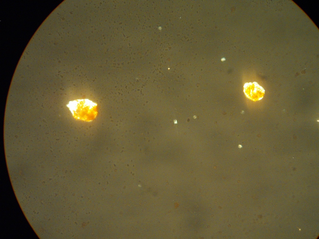 Cuthill Ferrihydrite Digital photograph Cross-Polarised Light (XPL) x1000, Field of view 0.1 mm
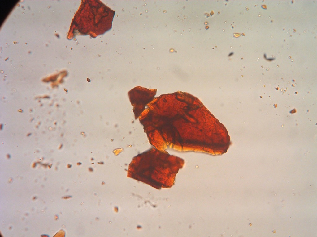 Saltburn, Ferrihydrite/Goetite Digital photograph Cross-Polarised Light (XPL) x1000, Field of view 0.1 mm
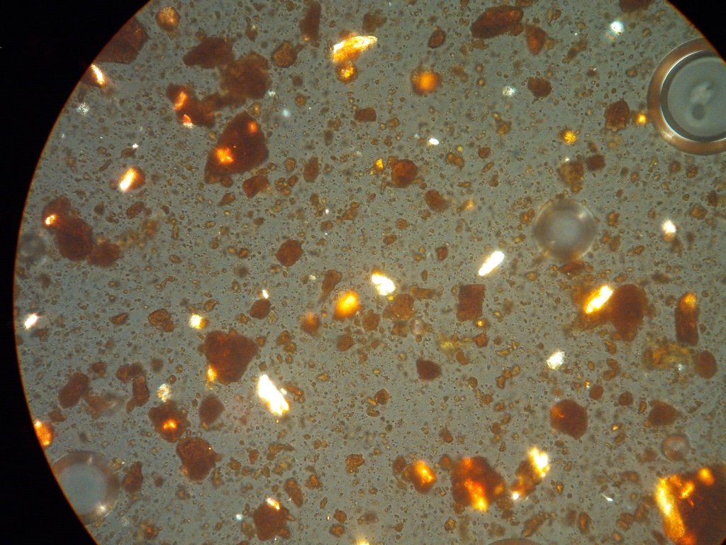 Six Bells, Ferrihydrite Digital photograph Cross-Polarised Light (XPL) x1000, Field of view 0.1 mm
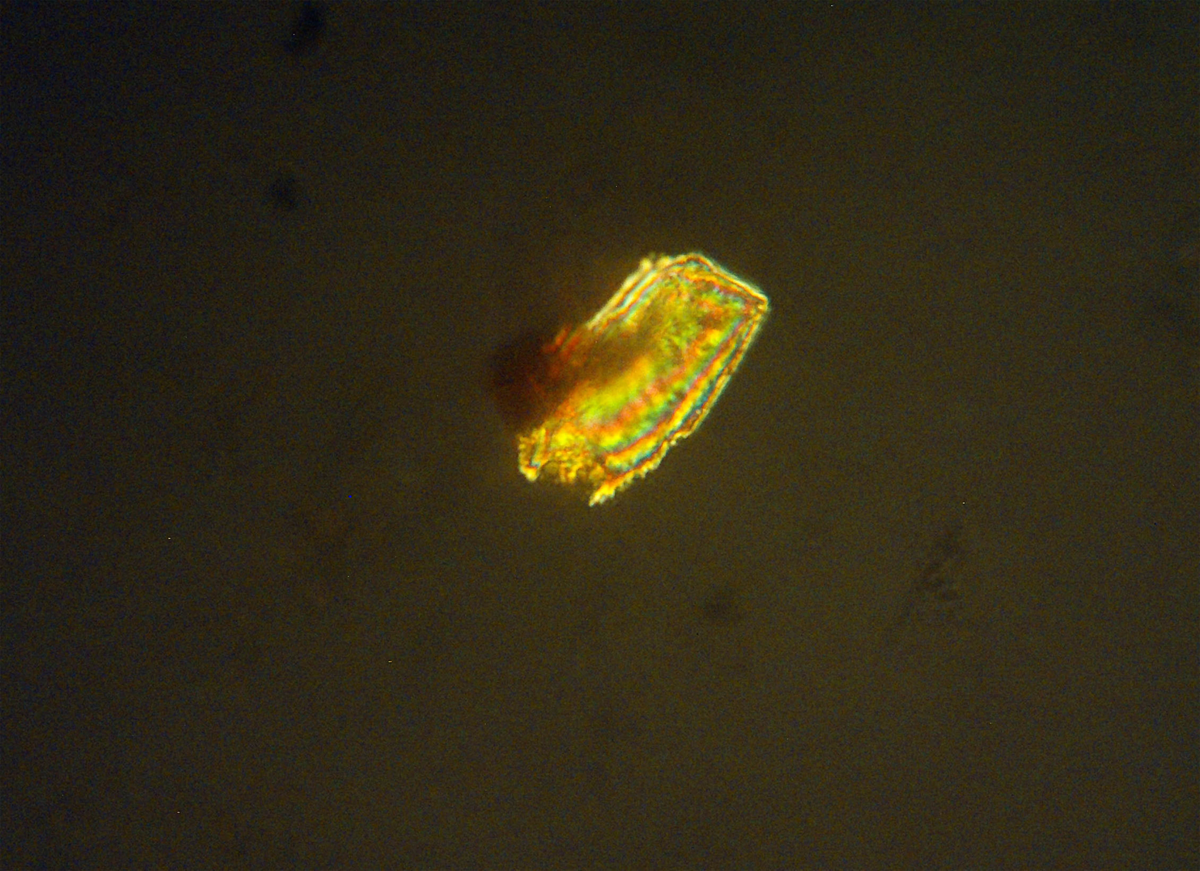
Six Bells Goetite Digital photograph Cross-Polarised Light (XPL) x1000, Field of view 0.1 mm
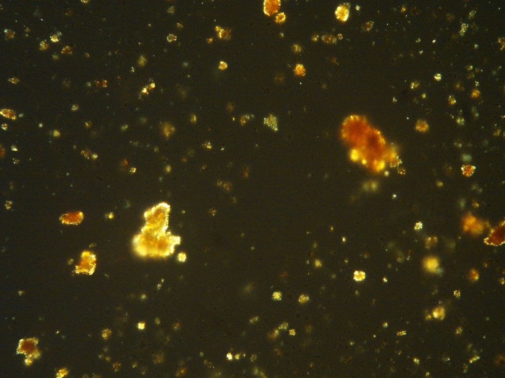 Tan-y-Garn Ferrihydrite Digital photograph Cross-Polarised Light (XPL) x1000, Field of view 0.1 mm
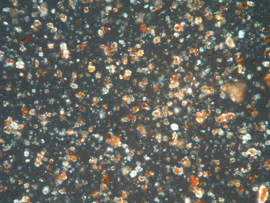
Tan-y-Garn, Ferrihydrite Digital photograph Cross-Polarised Light (XPL) x1000, Field of view 0.1 mm
The UCL Earth Science department were able to conduct X-ray diffraction tests on the pigments. A method that determines the atomic and molecular structure of a crystal by measuring the reflection patterns of a beam of X-rays off the crystal to determine its structure. The resulting graph reveals peaks according to where the beams have found a particular atomic spacing in a crystal structure. A small quantity of pigment, about a heaped teaspoon, was carefully placed in a crucible which was mounted and secured onto a rotating platform surrounded by a huge steel casing.
The results gave a clear indication of the presence of the crystalline structure of the mineral goethite. It was interesting to see this, and to know the exact mineral I was working with - to name it. Subsequently I have learned through handling the pigment - and comparing it to other similar goethite from established pigment sources - that its colour, texture and behaviour all consistently point to the mineral goethite.